The Body VR: Anatomy Viewer [Unreleased]
DICOM import, scaling, rotating, crop, annotation
Slicing, opacity tool, cross sectional viewing
DICOM segmentation, high-resolution anatomical dissections
DICOM import, Rotating, Scaling, Multi-planer visualization
Multi-modality of DICOM/MRI, Interoperability with any PACS system
Volumetric medical imaging, contouring, interactable LINAC model
Patient-specific VR reconstructions, 3D interaction
Segmentation and visualization, scaling, rotation
AR, Gaze based control, anchored model, clipping
3D visualization, Data cccess directly from PACS
Papers
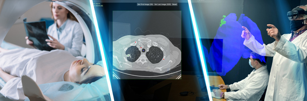
Applications of Virtual and Augmented Reality in Biomedical Imaging
Automated Segmentation, AR/VR, DICOM, radiological imaging, CT
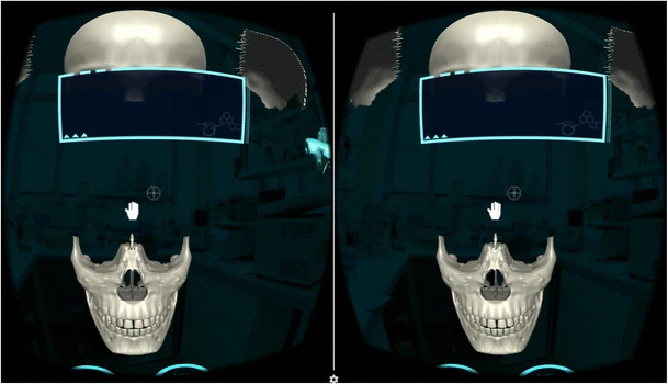
Virtual Reality Educational Tool for Human Anatomy
Educational, Interactive 3D visualization, VR, computed tomography
The Body VR: Anatomy Viewer [Unreleased]
Country US
Developer The Body VR LLC
Platform Oculus Rift, HTC Vive, Value Index
Commercial Availability N/A
Links https://store.steampowered.com/app/579620/The_Body_VR_
Keywords Medical imaging, DICOM, CT/MRI scan
Features DICOM import, scaling, rotating, crop, annotation
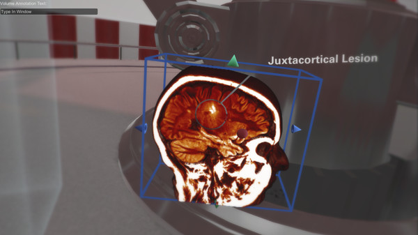
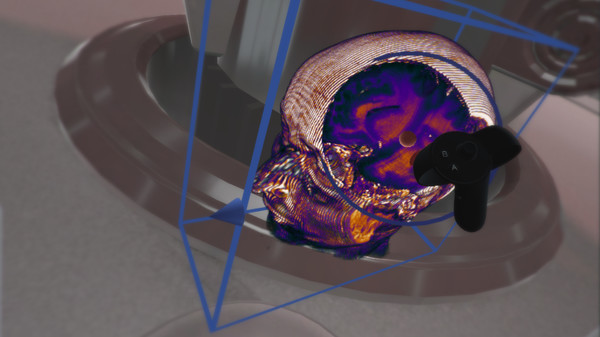
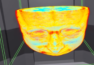
The Body VR: Anatomy Viewer displays 3D volumes generated from traditional CT/MRI scans that allows radiologists, medical students, and patients to view anatomical scans in virtual reality. The application includes scans from the brain, abdomen, heart, lungs, spine, and allows users to import their own DICOM files.
Users are able to directly interact with these models using hand controls, allowing a level of engagement on par with high end virtual reality applications. These controllers will allow the user to scale, rotate, and crop scans freely in 3D space. Users can add annotations to specific areas within a scan, to indicate lesions and tumors. The volumes can then be saved and uploaded to be viewed anywhere in the world, all patient health information is removed in the process. Users can select between multiple preset color schemes and even create your own. There are multiple environments built into the Anatomy Viewer to suit your preference.
(Text adapted from https://store.steampowered.com/app/579620/The_Body_VR_Anatomy_Viewer/)
ImmersiveView™ VR
Country US
Developer Immersive Touch
Platform Compatible With Major VR Platforms
Commercial Availability Oculus, VIVE
Links https://www.immersivetouch.com/immersiveview-vr
Keywords CT/MRI, Surgical planning, training, surgical simulations
Features Slicing, opacity tool, cross sectional viewing,
drawing and marking, plan and simulate surgical procedures
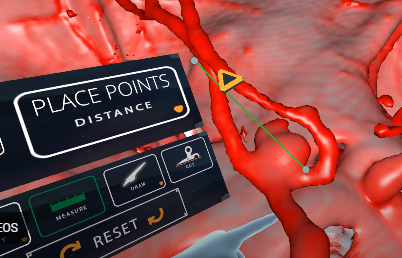
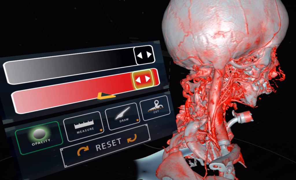
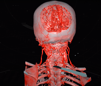
ImmersiveView™ VR converts CT, CBCT, 3D angiography and MRI scans into a ‘digital twin’ of the patient and provides a completely unique and unobstructed view of the case in virtual reality. No longer limited to certain angles of view, doctors can easily view the target anatomy, clearly and accurately, just as it exists in reality. Physicians handle the scan as if it were a physical object in the palm of their hand, observing from every angle to get the very best understanding of the situation, even flying into the anatomy to take a closer look. ImmersiveView™ VR provides unprecedented clarity in patient scans and is simple and intuitive to use. The platform is ideal for viewing critical scan information in all areas of the hospital: Radiology labs, ORs, ERs, and doctors’ offices.
Patients are imaged using common modalities like CT and MR. These scans are most frequently in a file format called DICOM. Under current processes, these patient DICOM scans are interpreted one slice at a time on a flat computer screen. The clinician must visualize anatomy and pathology by mentally “stitching” together the slides in their head.
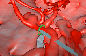
With ImmersiveView™ VR, 2D DICOM scans are merged and put into 3D virtual reality. Every conceivable angle of a patient’s unique anatomy can be engaged, from simple anatomy viewing to overlaying the original scan, creating measurements, placing virtual hardware, and much more.
(Text adapted from
https://www.immersivetouch.com/immersiveview-vr)
Further reading
- ImmersiveTouch Expands Availability of ImmersiveView™ Virtual Reality Solutions Into the Neurosurgery Market
- ImmersiveTouch Launches New Personalized Virtual Reality Imaging Platform into the Radiology Market
MEDICAL HOLODECK
Country Switzerland
Developer Medicalholodeck.com
Platform Valve Index, HTC Vive, Windows Mixed Reality
Commercial Availability Can be used with VR glasses from all manufacturers
Links https://www.medicalholodeck.com/en/
Keywords CT/MRI, DICOM, XR/VR
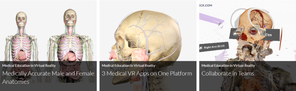
Features DICOM segmentation, high-resolution anatomical dissectionsl
Medicalholodeck’s virtual reality applications are used by medical professionals for surgery planning in teams and in medical education for teaching in virtual classrooms.
Medical Imaging XR is the DICOM Viewer for professional teamwork in virtual reality (VR). The software enables the import of medical image data from CT and MRI examinations in DICOM format and is used for surgery preparation and medical education. The app provides tools for editing, filtering, segmenting and measuring DICOM, OBJ and STL data in virtual reality and is ready for teamwork in VR. Groups of users can work together on DICOM and 3D data and discuss patient cases regardless of location. The app can be used with virtual reality glasses from all manufacturers.
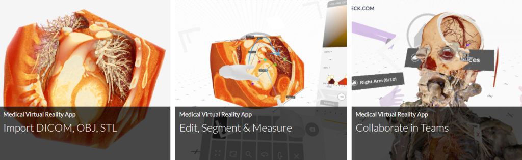
Dissection Master XR is the anatomy lab in virtual reality (VR). The app offers 3D data of human dissections in a VR classroom environment. It is ready for group teaching in virtual reality or by online streaming. The professionally dissected bodies are grouped and can be studied layer by layer. They are fully annotated and linked to Wikipedia for further information.
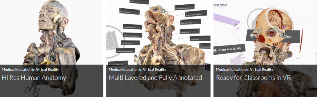
Anatomy Master XR Study the precise 3D male and female anatomical models of Zygote in VR. The high-resolution and carefully textured 3D models can be taken apart. They then convey the structure and organs of the human body as an interactive 3D object. The anatomical data are grouped. They can then be cut, filtered and enlarged in VR with various tools. Anatomy Master XR can be used simultaneously with Dissection Master XR and Medical Imaging XR. This enables the comparison of precise anatomical 3D models, medical patient data in DICOM format and high-resolution anatomical dissections. The software is ready for teamwork in VR. Teams can work on anatomical 3D models in VR at the same time.
(Text adapted from
Further reading
Medical Imaging VR
Country US
Developer hublab
Platform Valve Index, HTC Vive, Oculus Rift
Commercial Availability N/A
Links https://store.steampowered.com/app/1471980/MedicalImagingVR/
Keywords MRI, medical data, DICOM
Features DICOM import, Rotating, Scaling, Multi-planer visualization
Magnetic resonance imaging (MRI) is a type of scan that uses a strong magnetic field and radio waves to create detailed images of the organs and tissues within the body. An MRI scan uses a large magnet, radio waves, and a computer to create a detailed, cross-sectional image of internal organs and structures. This application allows to display different MRI scans and to browse into the different slices through a virtual reality environment. Users can also import their own MRI data (images from DICOM files) into the application to visualize them in VR.
(Text adapted from
https://store.steampowered.com/app/1471980/MedicalImagingVR/)

True 3D
Country US
Developer EchoPixel Inc
Platform
Commercial Availability N/A
Links https://echopixeltech.com/applications.html
Keywords MRI, DICOM
Features Multi-modality of DICOM/MRI, Interoperability with any PACS system
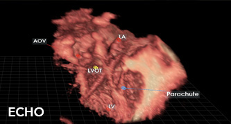
True 3D is a software platform that enables physicians to interact with virtual patient specific anatomy without the need for a bulky VR/AR headset. True 3D is used to assist physicians to plan surgical and interventional procedures to treat Congenital Heart Defects.
Patients with congenital heart defects and previous operations represent a visualization and surgical planning challenge due to a wide variation in morphologic features and clinical presentations. True 3D can provide 3D visualization for surgical interventions for a number of congenital heart defects.
(Text adapted from
https://echopixeltech.com/applications.html)
Further reading
The SuRgical Planner (SRP)
Country US
Developer Surgical theater
Platform
Commercial Availability Oculus, VIVE
Links https://surgicaltheater.net/services/
Keywords CT/MRI scans, surgical planning
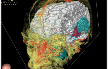
Features Patient-specific VR reconstructions, 3D interaction
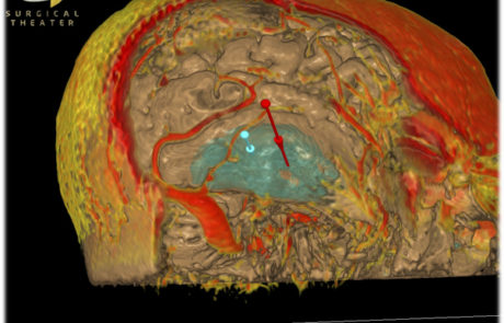
Surgical Planner accesses files from a patient’s traditional image modalities, such as CT or MRI scans, and processes the information to create patient-specific, VR reconstructions to help a neurosurgeon plan surgery, and educate the patient about their neurological condition.The interactive, immersive reconstruction can be seen on a touchscreen and through the use of a VR headset, such as Oculus Rift® or HTC Vive®. Surgeons can fly through the brain and locate medical issues such as a tumor or an aneurysm and study it from every angle with our medical virtual reality services. What SRP provides is especially important when developing an intricate surgical plan by allowing surgeons to create the best surgical approach based on the patient-specific VR reconstruction.
(Text adapted from
https://surgicaltheater.net/services/)
Further reading
- Surgical Theater Debuts a New Approach to Patient Engagement Utilizing Augmented Reality with Magic Leap
- Media and events of Surgical Theater
- http://www.bestaneurosim.com/simulator-surgical-theater/
DICOM VR
Country US
Developer Chris Williams (medical physicist)
and Kos Kovtun (radiation oncologist)
Platform
Commercial Availability N/A
Links http://www.dicomvr.com/
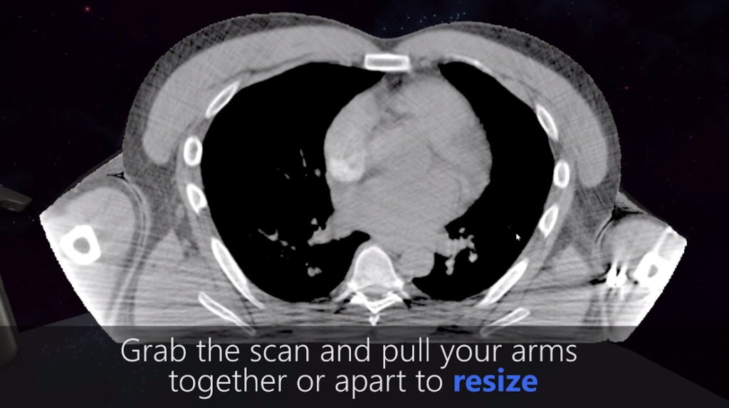
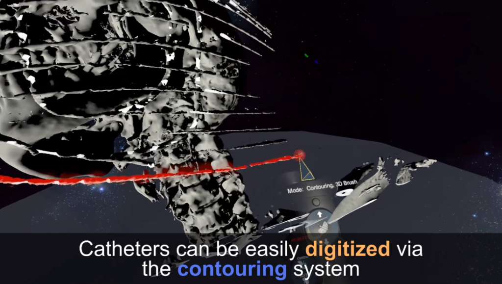
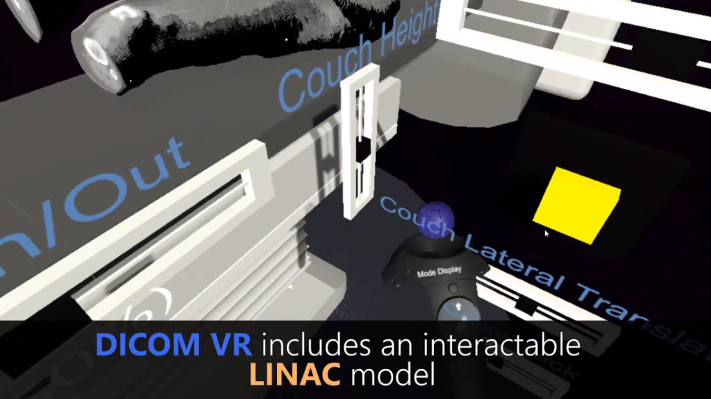
Keywords Volumetric medical imaging, DICOM
Features Volume rendering, clipping, different display modes,
contouring using 2D/3D brush, Interactable LINAC model
Chris Williams (medical physicist) and Kos Kovtun (radiation oncologist) work at the Brigham and Women’s Hospital/Dana Farber Cancer Institute. They are developing DICOM VR because of fundamental need to work with the increasing complexity of volumetric medical imaging. This work has recently been funded by a Brigham Research Institute Shark Tank grant.
(Text adapted from
Intravision XR
Country US
Developer DICOMDIRECTOR
Platform
Commercial Availability N/A
Links https://www.dicomdirector.com/intravision-xr/
Keywords 3D surgical modeling, DICOM, CT/MRI
Features Segmentation and visualization, scaling, rotation,
and plane on any device
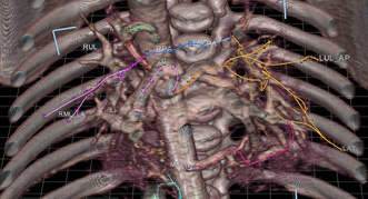
INTRAVISION XR is a cloud-based, 3D model medical software tool that automates the process of creating 3D models and allows you to visualize them. Fully formed 3D models are created from any DICOM dataset such as CT and MRI scans. Models contain complete anatomical detail, including spatial relationships between organs and their internal structure. View organ vasculature, neoplasms and anomalies in-situ within specific structures. Models are viewed in unparalleled visual detail using AR, VR or standard screens (phone, tablet, computer). Enhanced viewing of dimensional models in Augmented or Virtual Reality is the most realistic way to view anatomical structures.
(Text adapted from
https://www.dicomdirector.com/intravision-xr/)
Further reading
- How DICOM Director’s Intravision XR
uses Azure to power their groundbreaking HoloLens healthcare application - https://www.dicomdirector.com/learning-center/?_new_facet=intravision-xr
CommandEP
Country US
Developer SentiAR
Platform
Commercial Availability N/A
Links https://sentiar.com/commandep/
Keywords AR, 3D electroanatomic mapping
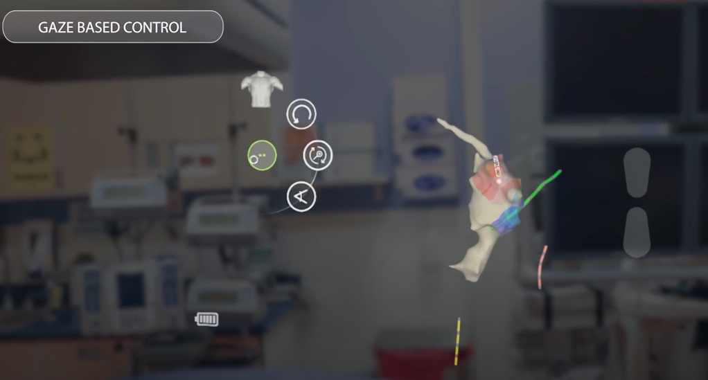
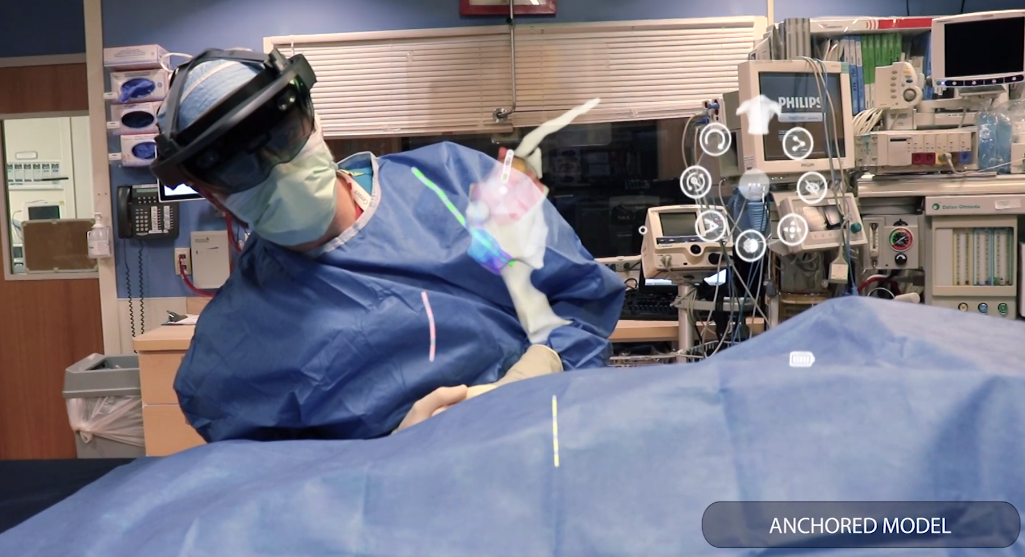
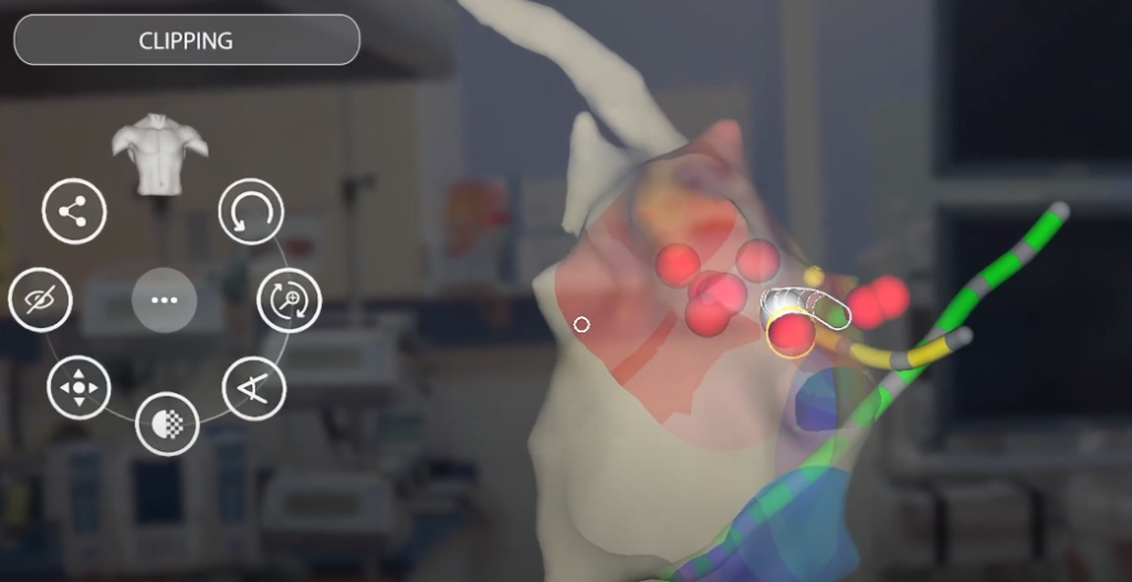
Features Gaze based control, anchored model, clipping
CommandEP uses real-time data from catheters and 3D electroanatomic mapping systems, to recreate an anatomical and electrical map of the particular patient’s heart.
(Text adapted from
https://sentiar.com/commandep/)
Further reading
- SentiAR Announces FDA 510K Clearance of CommandEP™ System, the First Holographic Cardiac Ablation Guidance System
- https://sentiar.com/news/
DICOM AI 3D VR VIEWER
Country US
Developer inlu
Platform PC + HTC Vive
Commercial Availability N/A
Links https://inlu.net/vr-projects/dicom-ai-3d-vr-viewer/
Keywords DICOM
Features 3D visualization, Data access directly from PACS
The initial need for the product appeared from a private medical institution in order to provide their patients with a truly visual and simply understandable way of their health condition. Specifically – DICOM scans. These scans are used widely in medical institutions around the world in order to present doctors with the inner body scan of the patient – a precise organ. This product virtualizes DICOM scans in order to present 3D images to doctors. They can then examine the model of the human body in full 3D mode, not only on a personal desktop but also via virtual reality glasses. Product enables in-depth exploration of organ condition and past changes examination along with patients.
(Text adapted from
https://inlu.net/vr-projects/dicom-ai-3d-vr-viewer/)
Papers
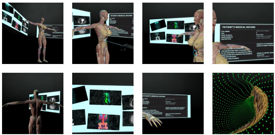
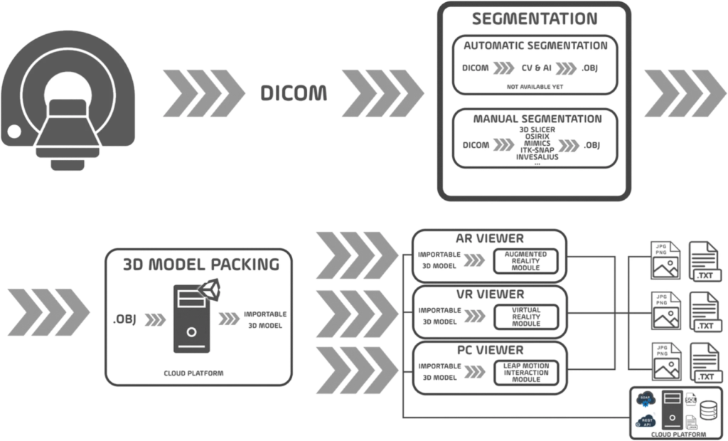
Applications of Virtual and Augmented Reality in Biomedical Imaging
Citation
González Izard, S., Juanes Méndez, J.A., Ruisoto Palomera, P. et al. Applications of Virtual and Augmented Reality in Biomedical Imaging. J Med Syst 43, 102 (2019). https://doi.org/10.1007/s10916-019-1239-z
Abstract
Virtual and Augmented Reality has experienced a steady growth in medicine in recent years. At the same time, the radiological images play a central role in the diagnosis and planification of surgical approaches. The aim of this study is to present the first attempt to enhanced radiological image visualization using virtual and augmented reality for better planification and monitorization of surgeries. This application allows to move beyond traditional two-dimensional images towards three-dimensional models that can be visualized and manipulated with both Augmented Reality and Virtual Reality. We propose possible approaches to automate the segmentation of radiological images, using computer vision techniques and Artificial Intelligence.
Keywords
Automated Segmentation, 3D visualization, AR/VR, DICOM, radiological imaging, CT
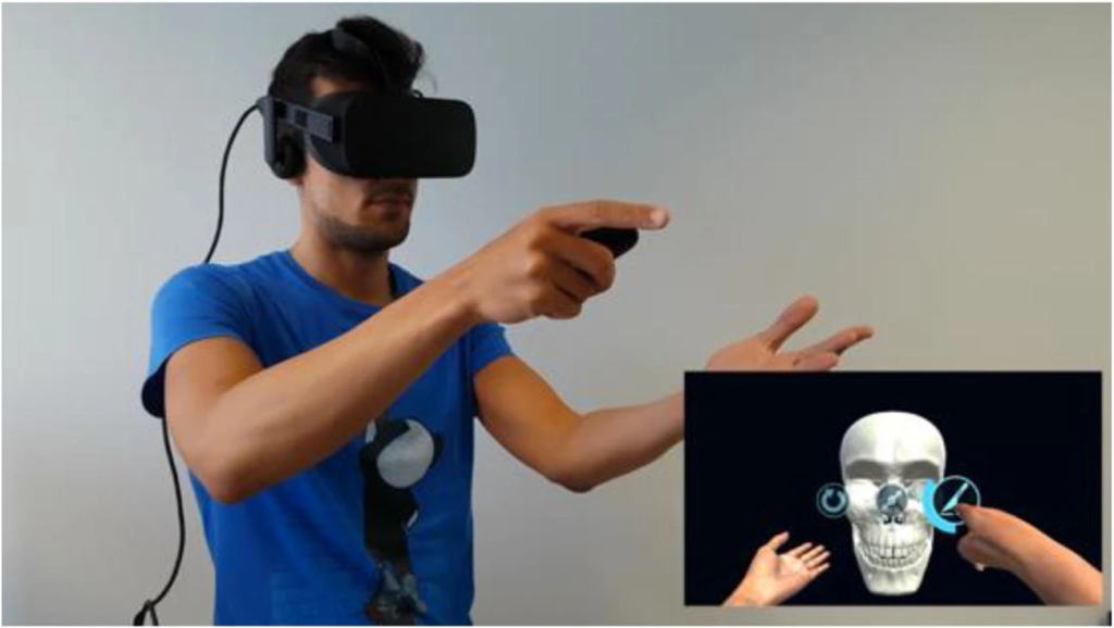
Virtual Reality Educational Tool for Human Anatomy
Citation
Izard, S.G., Juanes Méndez, J.A. & Palomera, P.R. Virtual Reality Educational Tool for Human Anatomy. J Med Syst 41, 76 (2017). https://doi.org/10.1007/s10916-017-0723-6
Abstract
Virtual Reality is becoming widespread in our society within very different areas, from industry to entertainment. It has many advantages in education as well, since it allows visualizing almost any object or going anywhere in a unique way. We will be focusing on medical education, and more specifically anatomy, where its use is especially interesting because it allows studying any structure of the human body by placing the user inside each one. By allowing virtual immersion in a body structure such as the interior of the cranium, stereoscopic vision goggles make these innovative teaching technologies a powerful tool for training in all areas of health sciences. The aim of this study is to illustrate the teaching potential of applying Virtual Reality in the field of human anatomy, where it can be used as a tool for education in medicine. A Virtual Reality Software was developed as an educational tool. This technological procedure is based entirely on software which will run in stereoscopic goggles to give users the sensation of being in a virtual environment, clearly showing the different bones and foramina which make up the cranium, and accompanied by audio explanations. Throughout the results the structure of the cranium is described in detailed from both inside and out. Importance of an exhaustive morphological knowledge of cranial fossae is further discussed. Application for the design of microsurgery is also commented.
Keywords
Educational, Interactive 3D visualization, VR, computed tomography